Aperio Digital Pathology Slide Scanners and Solutions
Scan, Manage, Analyze
Adopting digital pathology is complex, but it doesn’t have to be complicated. Leica Biosystems can clarify your organization’s path to digitally driven discoveries or diagnostics. Our world-leading, modular technology is supported by experience with thousands of Aperio Digital Pathology implementations worldwide.
当社の 利用規約 および プライバシーポリシー を事前にご確認の上、 お客さまのプライバシー上の選択についてもご理解ください。 ライカはお客さまのメールアドレスを第三者に共有することはありません。 送信いただいた個人情報に対し、当社は営業やマーケティングを始め、 製品やサービスの提供を目的として、お客さまに電話、Eメール、テキストメッセージなどを用いてご連絡をいたします。
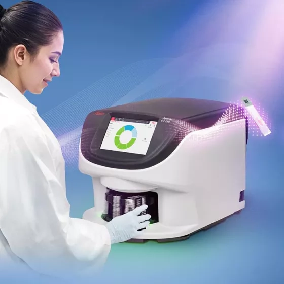
Introducing the new Aperio GT Elite™ scanner

Time is the New Currency
SPEED + INTELLIGENCE + CERTAINTY
Modern pathology demands more than speed alone. As workloads grow and expectations rise, speed must be matched with intelligence and certainty.
The Aperio GT Elite™ scanner brings all three together in one system, accelerating throughput while maintaining uncompromising standards, reducing manual touchpoints, and delivering dependable first-pass results that keep your lab moving with confidence at scale.

Confidence Comes From Consistency
The Aperio GT Elite scanner is engineered for first-pass scanning confidence across diverse specimens, stains, and protocols. Reliable image quality reduces rescans and protects both time and outcomes as volume increases. Optional Aperio iQC software further strengthens certainty by flagging and correcting for image quality issues during scanning, helping ensure results remain consistent even at very high throughput.

Intelligence that Works without Demanding Attention
Powered by SmartScan™ Technology, Certainty allows speed to scale without compromise. By internalizing workflow complexity, automatically calibrating each slide, optimizing scan parameters, and maintaining continuous, interruption free operation, the system reduces opportunities for human error and minimizes hands on involvement.

Speed is only Valuable when it can be Maintained
The Aperio GT Elite scanner is designed for high‑throughput scanning that holds performance hour after hour, delivering scans in 22 seconds per slide and up to 103 slides per hour. Optimized mechanics, efficient scan paths, and two‑way slide scanning work together to maximize output while delivering exceptional image quality. The result is consistent momentum across the lab. Speed that works under pressure.
Research Applications

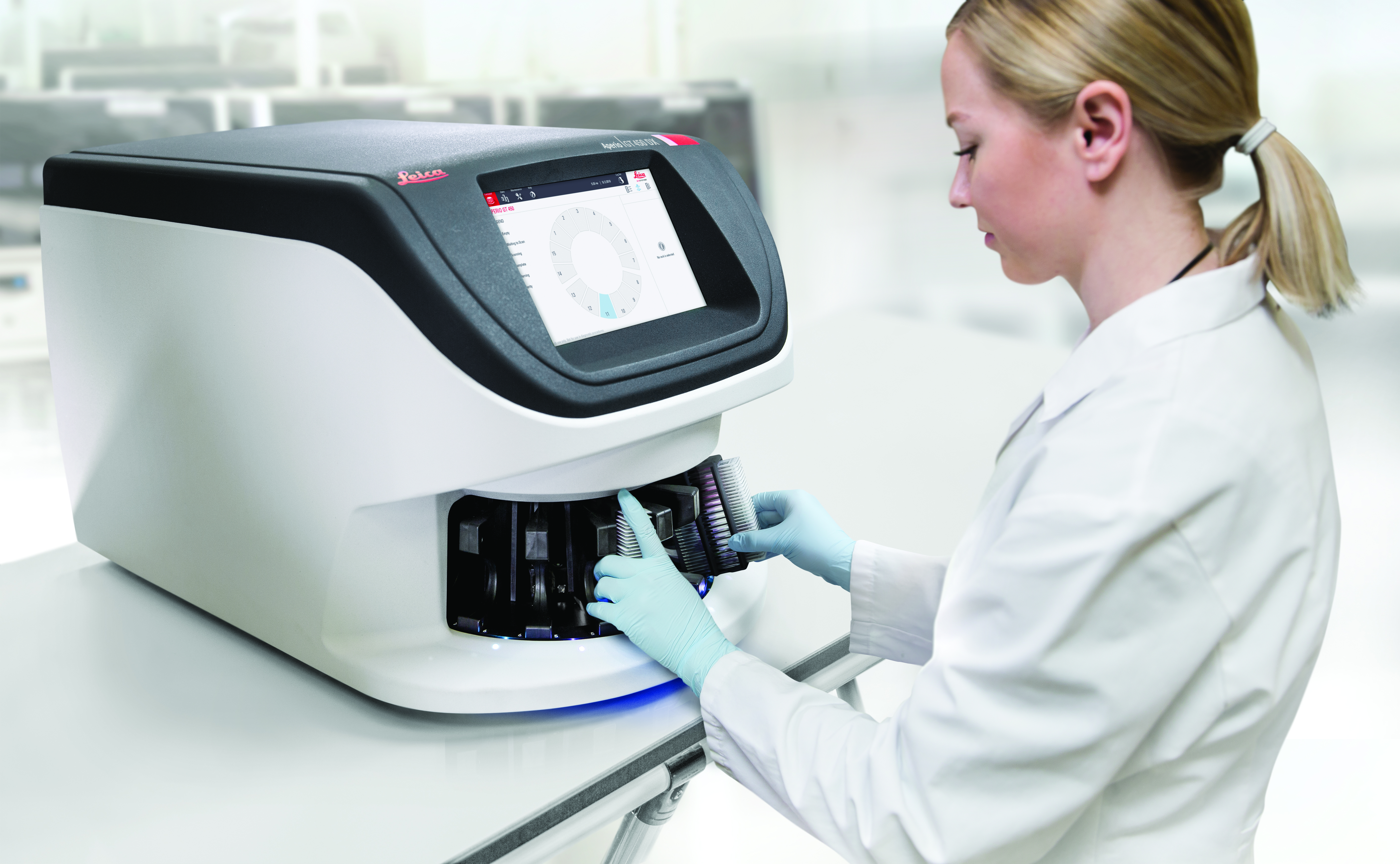
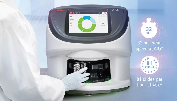
Aperio GT 450 scanner - Proven Technology for Researchers
With over 25 years of Digital Pathology innovation, Leica Biosystems delivers performance and reliability.
Our renowned Aperio GT 450 scanner has 40+ patents designed to support flexible scanning requirements and automated workflows. With a 32 second scan speed* and output of 81 slides per hour at 40x*, scanning can be completed quickly with confidence.
Continuous innovation, delivering scalable features,** including Manual Scan capabilities, DICOM compatible files, Extended Focus, and Aperio iQC software, provide efficient workflows and excellent image quality, ensuring seamless integration and secure, optimized delivery of your research.
*Scan speed assumes 15mm x 15mm area at 40x
** Optional features available to meet your workflow requirements. Some features not available in all countries.
Aperio GT 180 Scanner
With its mid-size slide capacity and advanced features, the Aperio GT 180 delivers the proven image quality and speed of trusted Aperio scanners in a design that works for your mid-range lab.
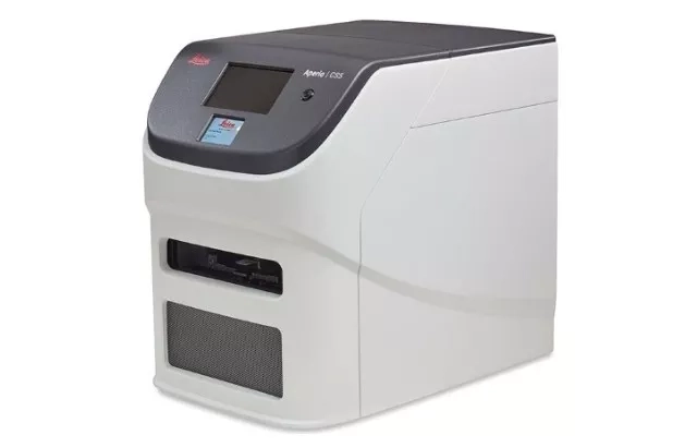
Aperio CS5 Scanner
Built to accommodate both 1×3 inch and 2×3 inch slides, the Aperio CS5 scanner delivers high-quality digital images in DICOM or SVS formats, with optional Z-stacking and 20X or 40X magnification. Its intuitive workflow makes it an excellent fit for research teams generating consistent, reproducible slide-level data across a wide range of studies.

Aperio FL Scanning Systems
The Aperio FL 120 slide scanner provides high-quality, high-resolution whole slide images of your research slides. Leverage the flexibility of brightfield, fluorescence, and FISH scanning in a single platform. For lower throughput needs, choose the 10-slide capacity Aperio FL 10.
Clinical Applications
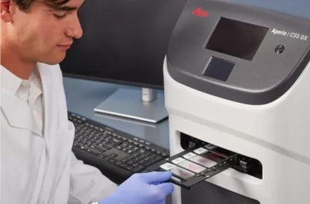
Aperio GT 450 DX scanner
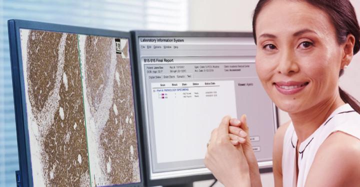
Aperio GT 450 DX scanner, WebViewer DX, eSlide Manager and LIS Connectivity
The Aperio GT 450 DX uses innovative color calibration technology to output consistent, high-quality images, enabling on-time, on-screen diagnosis. Aperio GT 450 DX is an automated, high-capacity digital pathology slide scanner that offers exceptional image quality, rapid delivery of cases, and high-performance, streamlined digital viewing built with the pathologist in mind.
HALO AP Dx software combined with the Aperio GT 450 DX scanner provides a complete, best-in-class clinical system for users leveraging digital pathology to advance and accelerate diagnostics.
HALO AP Dx (K252762) has received FDA 510(k) clearance for primary diagnostic use with the Leica Biosystems’ Aperio GT 450 DX Scanner in the USA. In addition, HALO AP Dx provides built-in compliance with FDA 21 CFR Part 11 and HIPAA.
Aperio CS5 DX scanner
Aperio CS5 DX scanner delivers a compact, cost-effective solution for low- to medium-volume labs, enabling seamless digitization of slides with exceptional image quality and minimal hands-on time. Its flexibility to handle both 1x3 inch and 2x3 inch slides, combined with manual scanning for complex cases, ensures adaptability across diverse workflows.
Aperio GT 180 DX scanner
The Aperio GT 180 DX scanner delivers the excellent image quality and speed of trusted Aperio scanners in a design that works for your clinical laboratory
Aperio GT 450 DX scanner
The Aperio GT 450 DX uses innovative color calibration technology to output consistent, high-quality images, enabling on-time, on-screen diagnosis. Aperio GT 450 DX is an automated, high-capacity digital pathology slide scanner that offers exceptional image quality, rapid delivery of cases, and high-performance, streamlined digital viewing built with the pathologist in mind.
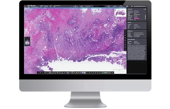
Software Solutions
Aperio Digital Pathology Software
Scalable. Accessible. Secure.
Effective implementation of digital pathology requires efficient management of images and data. Leverage Leica Biosystems' extensive pathology, workflow, and information technology expertise to integrate digital pathology in your organization.
Whether supporting a single institute installation or a multi-site hub and spoke architecture, our range of web-based software solutions provide an enterprise-level platform for your needs. Flexible deployment options facilitate on-premise, hosted, and cloud-based architectures, so you can choose the solution that is right for you.
For Research Use Only. Not for use in diagnostic procedures.

HALO Link Image Management System
HALO Link is a browser-based digital pathology software solution that helps research organizations worldwide to safely and securely manage, share, and analyze digital slides and data. Offering flexible data organization with robust search capabilities, seamless collaboration and custom integrations, HALO Link from Indica labs is a pathology workspace without bounds.
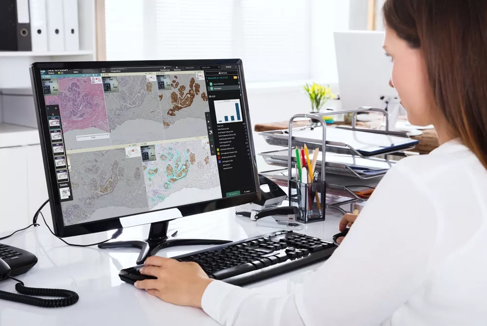
Aperio HALO AP Image Management System
An AI-powered, pathologist-centric image management system, Aperio HALO AP is designed for efficient on-screen slide review. It unifies data, whole slide images, and AI-driven analytics into a single, intuitive interface.
Aperio iQC Software
Seamlessly integrated with the Aperio GT series of scanners, Aperio iQC software enables rapid detection of six common artifacts and triggers alerts to the scanner console while slides are still on the scanner, ensuring that quality control is embedded directly into the scanning workflow. With intelligent automation, Aperio iQC ensures consistent artifact detection and categorization across tissue types and staining protocols
