The Impact of Decalcification on Staining
April 19, 2018 12:00pm CST
Different types of cancers frequently metastase to bone tissue. Treatment planning decisions are often based upon histology and special staining of these distant sites of disease. These decisions may rely on the outcome of immunohistochemistry, in situ techniques or molecular testing. Decalcification of the bone tissue is required to get quality histological slides. There are many decalcification reagents on the market. This presentation discusses the pros and cons of decalcification methods and how the laboratory should handle these specimens to best help the patient.
- Review the common decalcification methods
- Discuss the downstream effects of decalcification methods on staining.
- Describe the process of validation and reporting.
|
|
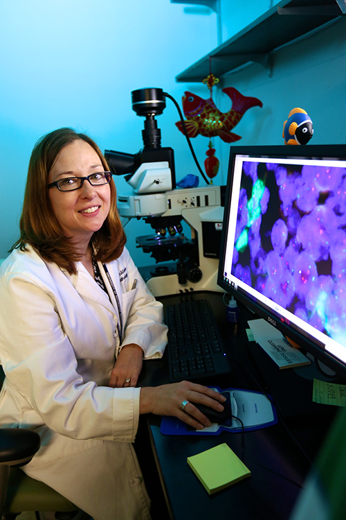
Loralee McMahon is the Immunohistochemistry Supervisor in Surgical Pathology at the University of Rochester Medical Center, Rochester, NY. She has worked as a histotechnologist for about 18 years. Her previous job was the Histology Supervisor at a Dermatopathology Lab in the DC area. Previous positions in histology included doing general histology and advanced staining for Alzheimer’s disease and Orthopaedics research. |

ABOUT US
Leica Biosystems (LeicaBiosystems.com) is a global leader in workflow solutions and automation, integrating each step in the workflow from biopsy to diagnosis. Our mission of "Advancing Cancer Diagnostics, Improving Lives" is at the heart of our corporate culture. Our easy-to-use and consistently reliable offerings help improve workflow efficiency and diagnostic confidence.

