
Immunofluorescence & FISH Image Analysis - Register for your FREE Demo
 New from Aperio Digital Pathology come 3 exciting new image analysis algorithms for fluorescence research applications.
Whether you work in Immunofluorescence or FISH, we have the powerful, flexible tools that can help to automate and standardize your research. Referenced in over 300 peer-reviewed publications, Aperio Image Analysis tools are trusted by researchers around the world.
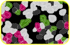 |
The Aperio Cellular IF Algorithm enables fast, accurate measurement and colocalization of up to 7 fluorescence channels, plus counterstain. It will distinguish between fluorphores localized within membrane, nuclear and/or cytoplasmic cellular compartments, in digital pathology whole slide tissue images. Retrieve objective, reproducible data from multiplex-stained tissue, which is challenging to quantitate by eye. With batch analysis options and seamless integration to Aperio eSlide Manager or Aperio Image Analysis Workstation, Aperio Cellular IF provides a flexible, powerful tool for your cellular immunofluorescence research needs. |
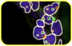 |
The Aperio FISH Brk/Fus algorithm enables highly-specific and sensitive detection of DNA sequence translocations (break-apart and fusion) in whole slide digital pathology images. It provides standardized results, removing the inter- and intra-user variability that is inherent in manual FISH counts. Aperio FISH Brk/Fus has been developed for full interoperability with the Aperio digital pathology solutions for researchers. It performs optimally on images captured with the Aperio VERSA or Ariol systems, and can be deployed through Aperio eSlide Manager or Aperio Image Analysis Workstation, depending on the throughput and unique needs of your organization. |
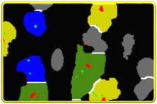 |
The Aperio FISH Amp/Del algorithm is designed to detect and quantify amplification and deletion of target gene sequences in digital pathology images generated from whole slide sections. It enables rapid analysis of all cells across a slide, and returns highly detailed and accurate signal counts without time-consuming manual effort. As the analysis is performed on digital images, there is no risk of probe fading, and no need to work in a dark room. Results are reproducible, and are permanently recorded in your Aperio eSlide Manager or Aperio Image Analysis Workstation database. |
ABOUT US
Leica Biosystems (LeicaBiosystems.com) is a global leader in workflow solutions and automation, integrating each step in the workflow from biopsy to diagnosis. Our mission of "Advancing Cancer Diagnostics, Improving Lives" is at the heart of our corporate culture. Our easy-to-use and consistently reliable offerings help improve workflow efficiency and diagnostic confidence.